Except for a recently discovered parasite of salmon, all animals require oxygen for survival and expel carbon dioxide as a waste product.1 Because animals are so varied, many breathing systems have evolved throughout the animal kingdom, each one designed to work in an animal’s particular environment. Depending on its size and habitat, an animal may employ direct diffusion, gills, lungs, or other respiratory passages for gas exchange.
In higher order animals, such as mammals, the respiratory and circulatory systems work together. In mammals, oxygen enters the body through the lungs where it attaches to hemoglobin in the blood and is then distributed by a matrix of vessels to cells and tissues. In the second half of the equation, carbon dioxide is collected from the cells and shunted back to the lungs where it is expelled by exhalation.
We’ve all seen pictures of tangled blood vessels that resemble the skeleton of a tree. The major vessels are thick like the trunk, and the branches start large and get progressively smaller as they stretch farther away. The vessels at the ends are downright tiny, but it’s these miniscule end pieces called capillaries that see all the action because that is where the gases are exchanged.
Right subject, wrong journal
Since this is the American Bee Journal — not the mammal journal — let’s take a look at the equivalent system in insects. Insects, too, have a respiratory and a circulatory system. Although the two systems work together to a limited extent, they remain separate. The respiratory system brings in oxygen and expels carbon dioxide while the circulatory system moves food in and waste out of the cells. But the insect circulatory system has no hemoglobin equivalent, so it has no ability to ferry oxygen from place to place throughout the body.
The parts needed to breathe
So how does a bug breathe without lungs or gills? In short, it has open ports on the integument that ultimately connect with the internal tissues, appendages, and organs. If you can envision an ocean liner with a series of portholes along its length, you’ve pretty much got the picture, except the bugs’ portholes allow air to enter 24/7. These holes, called spiracles, allow oxygen to move directly into the insect’s body.
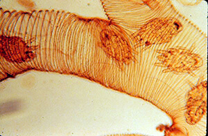
In bees, as in most insects, each spiracle opens into a tube called a trachea. The tracheae are formed from ingrowths of the integument and are circled with chitin rings, the same tough material that comprises the integument.2 Looking like a spring, the chitin spirals around the outside of each tracheal tube.
This spiral design — very similar to the coil-reinforced tubing used for vacuum cleaners, radiator hoses, and jack-in-the-boxes — makes the trachea flexible, stretchable, kink resistant, pressure proof, and abrasion immune. The strong, failsafe tubes can withstand all kinds of bee use and abuse.
From big to small
Just like the tree of blood vessels in mammals, the tracheae divide into smaller and smaller tubes as the distance from the spiracle increases. At the very end, where the tubes contact the cellular tissues, the tracheae become super tiny and are called tracheoles. The tracheoles, just like the corresponding capillaries in mammals, are the place where gas exchange takes place. The oxygen inside the tracheole diffuses into the tissue, and the carbon dioxide — which has been building up inside the cell — diffuses from the tissues for its trip back to the outside world.
Most insects have ten pairs of spiracles, two pairs in the thorax and eight in the abdomen. In bees, however, the first three pairs appear on the thorax and the remaining seven pairs line the abdomen. The spiracles are one of the first things you can see on a developing larva, visible as black dots along each side of the larva’s body. Soon after the spiracles become visible, you can also see a network of tracheae beginning to form, looking like a white-on-white roadmap.
Balloon-like air sacs
Instead of lungs, honey bees have thin-walled air sacs at various places along the tracheae. The sacs are located throughout the bee’s body, including the head, thorax, abdomen, and legs. Those in the abdomen are especially large, while the others are smaller.
These sacs, similar to balloons or pillows, expand or contract along with the bee’s need for oxygen. Changing pressure in the air sacs helps to move the oxygen to where it’s needed. Muscle movements in the abdomen control the flow of air in and out of the sacs. A bee can contract her muscles dorsoventrally (from top to bottom) or along the length of the abdomen (from front to back).
Because the muscles attach to the inside of the rigid abdomen, and not to the sacs directly, the entire abdomen moves as she pumps air through her tracheal system. When the abdomen contracts, air is squeezed from the sacs, and as the muscles relax, air is sucked into them, much like a bicycle pump.
The muscle contractions appear to flow along the length of the abdomen in an accordion-like manner, causing the abdominal plates to slide over one another. New beekeepers often become concerned when they observe these contractions for the first time, describing them as “waves of convulsions,” “muscle spasms,” or “seizures.”
The amplitude of the muscle contractions changes with the bee’s oxygen demand. Usually, the contractions are small and barely visible, while at other times they are pronounced. The amount of muscle movement can increase due to oxygen depletion or carbon dioxide elevation.3
The miracle spiracle
Pressure in the tracheal system could not increase without sealing the openings. Similar to a check valve on a water system, each spiracle lets ….


