
Recent validation of EFB test kits in North America could make the disease easier to diagnose
European foulbrood, or EFB, can be difficult to identify. Although the classical presentation — discolored larvae twisted like a corkscrew — is easy to recognize, there are many other ways the disease can manifest.
Larvae may have very little discoloration, creating a whitish, flaccid sac that looks a lot like an early case of chalkbrood. Larvae may succumb at many different ages, creating brown goop of various shapes and sizes, similar to what may occur from parasitic mite syndrome. Larvae may even succumb to the infection after being capped, when they are beginning to elongate into prepupae, yielding a sunken, sometimes perforated cap with a suspiciously American foulbrood (AFB)-like scale underneath.
Now, veterinarians are expected to be able to diagnose the disease as well. Thankfully, an EFB field-testing kit, using what’s called a “lateral flow device,” was developed over a decade ago, but it had not been validated in North America, until now.
Testing the test
“The lateral flow device is an incredibly useful tool, as long as it works,” says Dr. Meghan Milbrath, an assistant professor at Michigan State University. Milbrath and her colleagues recently evaluated these EFB test kits,1 which are manufactured by Vita Bee Health, and compared results against two other diagnostic methods: microscopy and PCR (“polymerase chain reaction,” a test which detects genetic material from EFB bacteria), both of which are very sensitive techniques. With growing evidence of EFB strain variation, which could allow the disease to escape detection, and little data on which strains are circulating, such validation is necessary to gauge the kit’s reliability.
“The results from the three methods seem to match very well for positive samples,” says Milbrath. “If a sample showed up positive for PCR, it also showed up positive for the lateral flow device, and showed up positive using the microscope.” While there were a handful of samples with inconsistent results, most of the time, data from the three methods agreed.
The researchers found that the kit’s sensitivity — or percent of colonies testing positive by the kit which also tested positive by PCR (the gold standard) — was 89.5%, whereas the sensitivity of microscopy was 97.4%. This means that about nine out of ten times, given a suspicious larval sample, the field-testing kit correctly detected EFB. Microscopy scored even higher, but the technique requires specialized equipment and training.
“If a beekeeper suspects that they have European foulbrood, it’s nice to know that they can go out, use the field test, and actually feel fairly confident in it when they get a positive result.” Milbrath recommends that beekeepers have both AFB and EFB test kits on hand, and notes that for the best results for EFB, care should be taken to select larvae showing only early signs of infection.
How it works
The field-testing kit uses the same lateral flow technology as COVID-19 rapid antigen tests. To use it, a biological sample (whether a discolored larva or a nasal swab) is thoroughly mixed in a bottle of liquid that comes with the kit, then a few drops of the liquid are dispensed onto a small sample well on the lateral flow cassette.
Absorbent material in the cassette wicks the liquid sample through a chamber containing antibodies linked to teeny tiny, colored particles. If the sample is infected, those antibodies will stick like Velcro to molecules (“antigens”) that come from the pathogen, and are pulled into the next chamber, where the whole works, including the colored particles, will stick to more antibodies that are fixed in place in a straight line.
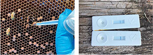
If the test worked properly and a blue line appears in the position labelled “T” (for “test”) in the viewing window, the sample is positive for EFB. Whether the test worked is indicated by another line at the “C” position (for “control”), which detects regular larval proteins that should be present in every sample. The test results can be read in just a few minutes.
These test kits cost somewhere around $15-16 per cassette, and can only be used once. That’s much cheaper than PCR diagnostics, without the hassle of shipping samples or long wait times for results. However, as Milbrath noted, there were a handful of colonies (6 out of 77) testing positive for EFB by PCR and microscopy, but negative according to the kit. This means that, although infrequent, the kits do come with the risk of false negative results.
At this point, it is not clear if the EFB in those six colonies escaped kit detection due to strain variation or some other problem with the kit. This will be an important point to distinguish, because if it is a problem with the kit, that could be overcome by using two tests per colony, whereas if strain variation makes the antigens unrecognizable by the kit’s antibodies, repeated tests won’t turn a false negative result positive. This kind of evasion of detection has been documented in Japan,2 but the prevalence of the offending strain elsewhere is not clear.
Milbrath and her colleagues are now sequencing the samples to see what EFB strains they harbor. Currently, the kit is manufactured using just one type of antibody against one specific EFB antigen, so, if strain variation is a widespread issue, new cassettes would have to be developed using multiple different antibodies to get around the problem.
In this study, the researchers did not evaluate the frequency of false positives — that is, given a healthy larva, how often did the test incorrectly say the larva had EFB? Given what we know about other rapid antigen tests, this is not likely to happen often if the kit is used correctly, and it would not depend on the EFB strain, like false negatives do. Despite this issue, a sensitivity of almost 90% is a big boost in confidence for the kit. And the results are virtually immediate, which lets you make management decisions right away, whereas by the time PCR results are reported back, it may be too late.
Peter Fowler, a veterinarian and graduate student at Michigan State who worked with Milbrath on the study, says that the microscopy method is quick but does require specialized equipment and knowledge. Typically, a small amount of larval sample is mixed with water on a cover slip, dried, stained with a specific dye, then inverted on a slide with a layer of oil and viewed under a microscope.
“It would be great if beekeepers could use this technique to help diagnose diseases, but it does take practice and training to differentiate some of these organisms,” Fowler says, adding that microscopy would be particularly beneficial for veterinarians to use to help ….